Vitamin E:Estrogen antagonist, energy promoter, and anti-inflammatory
Political scientists have recognized the process in which big corporations “capture” the governmental agencies that were created to regulate them. The editorial boards of professional journals can be captured even more cheaply than the agencies of government, and their influence can be even more valuable to industry.
If science impinges upon the plans of an industry, it can be managed into compliance, when the industry controls the journals and the agencies that fund research.
In the 1940s, it had already become clear to the estrogen industry that vitamin E research was impinging on its vital interests.
The Manhattan Project, that created the atomic bomb, also created a generation of scientific and bureaucratic zealots who ignored public health and safety to advance their projects and their careers, and changed the way science was done. At exactly the same time, the pharmaceutical industry was using its financial and political power to change the way medicine was practiced and taught, and the consequences for world health rivalled those of the nuclear industry.
In 1933 the physician R.J. Shute was aware of the problems associated with toxemia of pregnancy or preeclampsia. Especially among poorly nourished women, many pregnancies were complicated by circulatory problems, including cyclic bleeding, thrombosis, stroke, and hypertension, and these difficult pregnancies often ended in miscarriage or premature delivery, resulting in many serious health problems among the babies that survived.
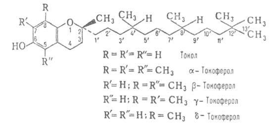
At that time, both estrogen and vitamin E were being widely studied, though the exact structure of the tocopherol molecule wasn’t defined until 1936-37. Vitamin E had been found to improve fertility of both male and female animals, and to prevent intrauterine death of the embryo or fetus, so it was called the “antisterility vitamin.” Using it to prevent women from having miscarriages must have occurred to many people.
Animal research in the 1930s was also showing that estrogen had many toxic effects, including causing infertility or intrauterine death, connective tissue abnormalities, and excessive blood clotting. Dr. Shute and his sons, Wilfred and Evan, were among those who considered vitamin E to be an antiestrogen. They found that it was very effective in preventing the clotting diseases of pregnancy.
Other researchers, who knew that progesterone protected against the toxic effects of estrogen, described vitamin E as the “progesterone-sparing agent,” since so many of its antiestrogenic effects resembled those of progesterone.
The Shute brothers began using vitamin E to treat circulatory diseases in general, rather than just in pregnant women–blood clots, phlebitis, hypertension, heart disease, and diabetes all responded well to treatment with large doses.
Vitamin E, as its name indicates, was the fifth type of “vitamin” factor to be identified, and it received its name in 1922, even though its chemical structure hadn’t been identified. The public quickly understood and accepted that certain substances in food were essential for life and health, so by 1940 practically all physicians were recommending the use of nutritional supplements.
If vitamin E was essential for human health, and achieved at least some of its amazing effects by opposing estrogen, then the synthetic estrogen industry had a problem.
Edward L. Bernays had already been in business for decades, teaching corporations and governments how to “engineer consent.” After his work for the government to engineer support for entering the first world war, Bernays’ next big job was for the tobacco industry. To convince women to smoke cigarettes, to achieve equality with men, he organized an Easter parade, Torches of Freedom, in which thousands of women marched smoking their freedom torches. In association with the American Medical Association (the editor of JAMA actually helped the tobacco industry design its campaigns), Bernays ran a campaign to convince Americans that smoking was good for the health.
The drug industry began using his techniques in sometimes crude but always effective ways. Estrogen was named “the female hormone;” natural hormones, including estrogen and progesterone, were claimed, without any research, to be inactive when taken orally. Physician-shills were created to claim wonderful effects for estrogen. The vitamin status of the tocopherols was denied; as recently as the 1970s (and maybe later), university professors of dietetics were flatly saying “no one needs vitamin E.”
Very little research showing the curative effects of vitamin E in human diseases was allowed to be published, so it was only occasionally necessary to openly denounce vitamin E as worthless or dangerous. In 1981, the journal of the AMA published an article reviewing the “toxic” effects of vitamin E. Since I had read all of the articles cited, I realized that the author was claiming that whenever vitamin E changed something, the change was harmful, even though the original publication had described the effect as beneficial.
Although JAMA was eventually forced to give up its revenue from cigarette advertising, it didn’t suffer at all, because of the vast advertising campaigns of the estrogen industry. JAMA obviously wouldn’t want to publish anything suggesting that vitamin E, or progesterone, or thyroid, might be beneficial because of its antagonism of the harmful effects of estrogen.
Estrogen causes changes in the uterus that prevent implantation of the embryo, and that impair support for its development if it has already implanted. It decreases the availability of oxygen to the embryo, while vitamin E increases it.
My dissertation adviser, A.L. Soderwall, did a series of experiments in which he showed that providing hamsters with extra vitamin E postponed the onset of infertility in middle age. In my experiments, vitamin E increased the amount of oxygen in the uterus, correcting an oxygen deficiency produced either by supplemental estrogen or by old age. Progesterone has similar effects on the delivery of oxygen to the uterus.
In the 1940s, the official definition of vitamin E’s activity was changed. Instead of its effectiveness in preventing the death and resorption of embryos, or the degeneration of the testicles or brain or muscles, it was redefined as an antioxidant, preventing the oxidation of unsaturated oils.
Although some people continued to see vitamin E as a protective factor in thrombosis, heart attacks, diabetes, and infertility, the medical establishment claimed that results in animal diseases couldn’t be applied to humans, and that a mere antioxidant couldn’t prevent or cure any human disease.
In the experiments that led to the discovery of vitamin E, rats were fed rancid lard, and were given cod liver oil as a source of vitamin A. Both of these contain large amounts of polyunsaturated oils.
Between 1929 and the middle 1930s, the nutritional essentiality of polyunsaturated fatty acids was claimed to have been demonstrated experimentally. Like the vitamin E experiments, these were done on rats, but this time the medical establishment didn’t argue that the requirement for linoleic or linolenic acid in humans hadn’t been proven by rat experiments, as they had argued regarding vitamin E. By the time a group of vitamin B6 researchers showed, in the 1940s, that the supposed “essential fatty acid deficiency” syndrome was cured by supplementing vitamin B6, the ability of polyunsaturated fatty acids to suppress the metabolic rate and to decrease all nutritional requirements had become clear. The “essential” fatty acids strongly inhibit the production of thyroid hormone.
When the results of these experiments are considered together, the striking fact is the toxicity of the polyunsaturated oils, suppressing metabolism in some experiments, and creating a variety of degenerative conditions in others.
In the late 1940s and early 1950s, estrogens of many types were being synthesized from hydrocarbons, and promoted for preventing miscarriages, because “estrogen is the female hormone.” The livestock industry found that adding polyunsaturated oils to animal food was very profitable, because it slowed metabolism and produced cheap weight gains; then the antithyroid oils were promoted as “heart-protective” for humans, though they promote both heart disease and cancer by suppressing the thyroid and destroying vitamin E. (The promotion of estrogen in animal feeding to increase the efficiency of fattening, and for humans as “preventing heart attacks,” made an interesting parallel with the campaigns for the oils.)
Under the influence of the food industry, researchers lost sight of the unsafety of the oils for food, and supported the idea that vitamin E is merely an antioxidant, and that seed oils are the best source of vitamin E.
At that time, there was no understanding that the antifertility effects of the polyunsaturated oils shown in the vitamin E research were related to the antifertility effects of estrogen. I think the key to understanding the nature of vitamin E is to understand the close interaction between estrogen and the polyunsaturated fatty acids (PUFA). Their mechanisms are closely intertwined, and are opposed by a variety of stabilizing and energizing substances, including saturated fats, progesterone, thyroid, vitamin E, and aspirin.
Chemicals that inhibit enzymes are generally toxic, producing a specific symptom or deterioration. But a group of enzymes associated with estrogen and PUFA is inhibited by the protective substances mentioned above. In our modern diet, these PUFA-metabolizing enzymes in the fetus and infant act on endogenous fats of the Mead acid type. Mead acid is anti-inflammatory and has a wide range of protective effects. The dietary PUFA interfere with the activity of these natural protective substances.
If we didn’t eat PUFA, these enzymes would regulate Mead acids, and when activated by stress would produce anti-stress substances to limit the stress reaction. But as we accumulate more anti-vitamin E fats, these enzymes, instead of stopping the inflammatory process, promote it, and produce tissue damage. Other stress-limiting factors, such as progesterone, tend to eliminate the conditions that activate these enzymes, in a very indirect kind of inhibition, by correcting the abnormalities caused by stress.
Estrogen increases events associated with inflammation, and vitamin E decreases them. Estrogen produces capillary leakiness, and vitamin E opposes it. Estrogen increases platelet aggregation and decreases the factor that inhibits platelet aggregation; vitamin E works in the opposite direction.
Excessive clotting is known to be caused by excessive estrogen, as well as by vitamin E deficiency.
Excessive clotting leads to fibrosis, and there is clear evidence that vitamin E prevents and cures fibrotic diseases, though powerful medical institutions still fail to recognize this evidence. Estrogen and polyunsaturated fats increase fibrosis.
Estrogen increases prostaglandin synthesis, vitamin E decreases it; estrogen increases COX and LOX enzymes, vitamin E decreases them. (Jiang, et al., 2000; Ali, et al., 1980; Parkhomets, et al., 2001). Estrogen releases enzymes from lysosomes, vitamin E inhibits their release. Beta-glucuronidase, one of these enzymes, can release estrogen at the site of inflammation.
Estrogen often increases intracellular calcium and protein kinase C, vitamin E opposes these effects.
Polyunsaturated fatty acids and their prostaglandin derivatives act as effectors or amplifiers of estrogen’s action.
If vitamin E acts as a protector against polyunsaturated fatty acids, that fact by itself will explain at least some of its antiestrogenic effects.
Besides opposing some of the downstream effects of the toxic fatty acids, vitamin E inhibits lipolysis, decreasing the concentration of free fatty acids (estrogen does the opposite), and it binds and inactivates free fatty acids. Its long saturated carbon chain is essential for its function, and this saturated chain may allow it to serve as a substitute for the omega-9 fats that produce Mead acid. The unsaturated tocotrienols have hardly been tested for true vitamin E activity, and in animal studies they were suggested to be toxic, causing enlargement of the liver.
One of the most important unstudied anti-PUFA effects of vitamin E is its direct destruction of linolenic and linoleic acids. Bacterial vitamin E is known to be involved in the saturation of unsaturated fatty acids, and it is known that intestinal bacteria convert linoleic and linolenic acids to the fully saturated stearic acid.
“No metabolic function is known for alpha-tocopherolquinol or its quinone other than as a cofactor in the biohydrogenation of unsaturated fatty acids that can be carried out by only a few organisms.”
— P. E. Hughes and S. B. Tove, 1982.
“Linoleic acid was significantly decreased (P < 0.001) and there was a significant rise (P < 0.05) in its hydrogenation product, stearic acid. Linolenic acid was also significantly decreased. … The study provides evidence that bacteria from the human colon can hydrogenate C18 essential polyunsaturated fatty acids.”
— F. A. Howard and C. Henderson, 1999.
Because the decision to view vitamin E as a simple antioxidant was politically determined, until recently no one was eager to emphasize the pathology of lipid peroxidation and free radicals, partly because lipid peroxidation is only a minor part of the toxicity of polyunsaturated oils, and research into the true nature of their toxicity receives little support. Under these circumstances, even the factual antioxidant parameters of various forms of vitamin E have been distorted. (See, for example, Chen, et al., 2002).
People who say vitamin E is nothing but an antioxidant sometimes take other antioxidants, along with or instead of vitamin E. It is common to hear that BHT, BHA, and a variety of natural compounds (derived from industrial and agricultural waste) are “better antioxidants than vitamin E.” Anything that can be oxidized and reduced (melatonin, estrogen, tryptophan, carotene, etc.) will function in some systems as an antioxidant, and under other conditions may become a pro-oxidant.
People who think of abstract “antioxidant” function as beneficial seem to visualize something like an omnipresent fire department, putting out each fire as it starts. I think it is more reasonable to think of biological antioxidant systems as arson prevention programs, preventing the fire from ever getting started.
Since the requirement for vitamin E decreases as consumption of unsaturated fats decreases, if we didn’t eat significant amounts of those fats, our requirement for it would be very small, if not zero.
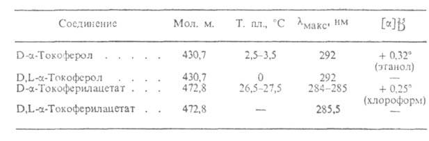
In the years since the tocopherols were identified as vitamin E, the material sold for research and supplementation has changed radically several times, even after it was specifically identified as mixed tocopherols or d-alpha tocopherol. Variations in viscosity and color, caused by changes in impurities, have undoubtedly influenced its biological properties, but the belief in its antioxidant nature has kept researchers from asking what a particular batch of vitamin E really consists of and what it actually does.
“We compared the effects of mixed tocopherol vs alpha-tocopherol alone on SOD activity and iNOS expression in cultured myocytes exposed to H-R. … Both tocopherol preparations attenuated cell injury… However, the mixed tocopherol preparation was significantly superior to alpha-tocopherol in myocyte protection… The lack of efficacy of commercial tocopherol preparations in clinical trials may be related to the absence of gamma- and delta-tocopherols.”
— Chen H, Li D, Saldeen T, Romeo F, Mehta JL, Biochem Biophys Res Commun 2002.
It is probably better to stay as far away from polyunsaturated fats in the diet as possible, to create the protective “deficiency” state seen in animal studies (against cancer, trauma, poisoning, shock, inflammation, infection, etc.), rather than trying to saturate oneself with antioxidants, given the poorly understood nature of vitamin E products and the known toxicity of many other antioxidants on the market.
The carcinogenic properties of polyunsaturated fats have been known for over 50 years, as has the method of life extension through dietary restriction. Recently, several studies have shown that long-lived species have less highly unsaturated fats than short-lived species. Calorie restriction prevents lipids in the brain, heart, and liver from becoming more unsaturated during aging. (Lee, et al., 1999; Laganiere, et al., 1993; Tacconi, et al., 1991; R. Patzelt-Wenczler, 1981.)
When cells are grown in tissue culture without “essential fatty acids,” they enter a state of “deficiency,” become extremely resistant to chemical injury, and can proliferate indefinitely. These experiments not only demonstrate how polyunsaturated fats sensitize cells to damage (Wey, et al., 1993), but should also embarrass those who base their arguments for the essentiality of these oils on their supposed requirement for “building cell membranes.” Since cells can multiply indefinitely in a state of “essential fatty acid deficiency,” we must conclude that these oils are not needed for “membranes,” or perhaps that “without membranes” cells resist damage better.
Conversely, excess insulin and prolactin or vitamin E deficiency increase the activity of enzymes that convert linoleic acid into more highly unsaturated fatty acids. Excess insulin and prolactin play important roles in many degenerative diseases.
Highly unsaturated fats suppress respiration through various pathways, and there is a tendency for unsaturation to increase with aging, endocrine stress, and vitamin E deficiency, in parallel with the decrease in respiratory energy production over the lifespan. Many studies show that vitamin E can protect and enhance mitochondrial energy production. (Kikuchi, et al., 1991; Donchenko, et al., 1990, 1983; Guarnieri, et al., 1981, 1982.) However, the state of so-called fatty acid deficiency not only makes mitochondria very resistant to damage, but also greatly enhances their energy production. Supplemental vitamin E is rarely as effective as the absence of toxic oils from the diet.
Many food composition tables no longer list liver as a good source of vitamin E, even though most of an animal’s vitamin E is stored in its liver. This bias in the nutrition literature can be traced to several sources, but the major influence was the 1970s campaign organized by pharmaceutical companies that had patented new forms of synthetic “vitamin A.” They had physicians and professors fabricate and place stories in national magazines about the severe toxicity of natural vitamin A, clearing the field for supposedly non-toxic new drugs, which turned out to be catastrophically toxic. As a result, many people, fearing vitamin A, stopped eating liver. Other liver vitamins, including vitamin K, work in close association with vitamin E, and stably stored forms of vitamin E are likely a good approximation for meeting our needs.
There remains a strong divide between what people can say in their professional publications and what they actually believe. The man under whose influence vitamin E was designated an antioxidant, M. K. Horwitt, expressed dismay when the government raised the recommended intake of vitamin E by 50%, because that decision was not supported by new data, and because millions of people receive only ten milligrams a day and are “healthy.” Yet he himself took 200 mg daily (plus aspirin) for many years. Apparently, he didn’t put much faith in the ideas he publicly promoted.
References
- Prostaglandins Med 1980 Feb;4(2):79-85. Inhibition of human platelet cyclooxygenase by alpha-tocopherol. Ali M, Gudbranson CG, McDonald JW. “Alpha-tocopherol, an inhibitor of platelet aggregation, was evaluated for its effects on the synthesis of thromboxane and prostaglandins. A dose-dependent reduction in thromboxane B2 and prostaglandin D2 synthesis was observed with approximately 60% inhibition at 5.0 IU or alpha-tocopherol. Alpha-tocopherol produced a time-dependent, irreversible inhibition.”
- Int J Vitam Nutr Res 2001 Jan;71(1):18-24. Vitamin E and the prevention of atherosclerosis. Bron D, Asmis R. “Recent new findings have shed new light on the physiological role of vitamin E and suggest that it has a much broader array of biological activities than originally expected. In addition to its well described role as an antioxidant, it is becoming evident that vitamin E also can modulate the immune system, suppress local and chronic inflammation, reduce blood coagulation and thrombus formation, and enhance cell function and survival.”
- Plast Reconstr Surg 1981 Nov;68(5):696-9. The effectiveness of alpha-tocopherol (vitamin E) in reducing the incidence of spherical contracture around breast implants. Baker JL Jr. “Vitamin E appears to be a safe, simple, and inexpensive means of reducing the number of postoperative capsular contractures following breast augmentation. The synthetic form of vitamin E (alpha-tocopherol) is recommended to avoid nausea or skin eruptions in patients with oily skin, which are frequently encountered when the natural form is taken. No harmful side effects have been noted in any of the patients to date. Vitamin E has no effect on coagulation systems and does not cause excessive bleeding either during or after surgery. The recommended dosage of synthetic vitamin E is 1000 IU, b.i.d., for 2 years beginning 1 week before surgery. If no contracture exists at that time, the dosage may be reduced to 1000 IU daily thereafter.”
- Carcinogenesis 1999 Jun;20(6):1019-24. Decrease in linoleic acid metabolites as a potential mechanism in cancer risk reduction by conjugated linoleic acid. Banni S, Angioni E, Casu V, Melis MP, Carta G, Corongiu FP, Thompson H, Ip C.
- Mech Ageing Dev 1978 Nov;8(5):311-28. Anomalous vitamin E effects in mitochondrial oxidative metabolism. Baumgartner WA, Hill VA, Wright ET. “Three different vitamin E effects, suggestive of specific antioxidant effects, were discovered in the protective action of vitamin E against respiratory decline (a decrease in mitochondrial respiration attributed to a ‘leakage’ of electron transport radicals). No correlation was found between respiratory decline and random lipid peroxidation. The mechanisms behind two of the three atypical vitamin E effects were defined. Both involve an artifact in the TBA assay for lipid peroxidation. This artifact occurs when TBA assays are carried out in the presence of sucrose and acetaldehyde; the latter is produced from ethanol, the solvent used to add vitamin E to preparations. The artifact in the TBA assay for peroxidations appears also to be responsible for differing interpretations of the hepatotoxic effect of ethanol.”
- Eur J Biochem 1990 Mar 10;188(2):327-32. Polychlorinated biphenyls increase fatty acid desaturation in the proliferating endoplasmic reticulum of pigeon and rat livers. Borlakoglu JT, Edwards-Webb JD, Dils RR.
- Nutr Cancer 2000;38(1):87-97. Effects of topical and oral vitamin E on pigmentation and skin cancer induced by ultraviolet irradiation in Skh:2 hairless mice. Burke KE, Clive J, Combs GF Jr, Commisso J, Keen CL, Nakamura RM. “Results showed that the skin concentrations of Eol, as well as levels in the adipose tissue, were increased after topical application. Mice treated with each form of vitamin E showed no signs of toxicity and had significantly less acute and chronic skin damage induced by UV irradiation, as indicated by reduced inflammation and pigmentation and by later onset and lesser incidence of skin cancer.”
- Am J Physiol 1991 Jun;260(6 Pt 2):R1235-40. Acute phase response in exercise. II. Associations between vitamin E, cytokines, and muscle proteolysis. Cannon JG, Meydani SN, Fielding RA, Fiatarone MA, Meydani M, Farhangmehr M, Orencole SF, Blumberg JB, Evans WJ.
- Vrach Delo 1990 Dec;(12):6-8. [The effect of tocopherol and nicotinic acid on the microcirculation and blood coagulability in patients with ischemic heart disease] Chernomorets NN, Kotlubei GV, Vatutin NT, Zhivotovskaia IA, Gnilitskaia VB, Alifanova RE, Lobach EIa, Mal’tseva NV, Mitrofanov AN. “Complex treatment using tocopherol acetate produced a positive effect on the coagulation properties of the blood and did essentially influence the fibrinolytic activity and microcirculation. Tocopherol plus nicotinic acid resulted in normalization of the blood coagulation process, favoured activation of fibrinolysis and improvement of the microcirculatory bed.”
- Free Radic Biol Med 1991;10(5):325-38. Oxidative status and oral contraceptive. Its relevance to platelet abnormalities and cardiovascular risk. Ciavatti M, Renaud S. INSERM Unit 63, Bron, France. “Oral contraceptive (OC) use is a risk for thrombogenic events. … From these data we conclude that: 1. OC use modifies slightly but significantly the oxidative status in women and in animals by decreasing in plasma and blood cells the antioxidant defenses (vitamins and enzymes). 2. The changes in the oxidative status are related to an increase in plasma lipid peroxides apparently responsible for the hyperaggregability and possibly the imbalance in clotting factors associated with the OC-induced prethrombotic state. 3. These effects of OC appear to be increased by a high intake of polyunsaturated fat and counteracted by supplements of vitamin E. 4. The risk factors acting synergistically with OC, have all been shown to increase platelet reactivity.”
- Bol Med Hosp Infant Mex 1980 May-Jun;37(3):457-67. [Jaundice caused by microangiopathic hemolysis associated to septicemia in the newborn] Covarrubias Espinoza G, Lepe Zuniga JL. “These infants with over% fragmented cells were found to have a significant association with: sepsis, jaundice, crenated RBC’s, low levels of hemoglobin, increased reticulocyte count, and low vitamin E levels.”
- Endocrinology 1992 Nov;131(5):2482-4. Vitamin E protects hypothalamic beta-endorphin neurons from estradiol neurotoxicity. Desjardins GC, Beaudet A, Schipper HM, Brawer JR. “Estradiol valerate (EV) treatment has been shown to result in the destruction of 60% of beta-endorphin neurons in the hypothalamic arcuate nucleus. Evidence suggests that the mechanism of EV-induced neurotoxicity involves the conversion of estradiol to catechol estrogen and subsequent oxidation to free radicals in local peroxidase-positive astrocytes. In this study, we examined whether treatment with the antioxidant, vitamin E, protects beta-endorphin neurons from the neurotoxic action of estradiol. Our results demonstrate that chronic vitamin E treatment prevents the decrement in hypothalamic beta-endorphin concentrations resulting from arcuate beta-endorphin cell loss, suggesting that the latter is mediated by free radicals. Vitamin E treatment also prevented the onset of persistent vaginal cornification and polycystic ovarian condition which have been shown to result from the EV-induced hypothalamic pathology.”
- Free Radic Biol Med 2000 Dec 15;29(12):1302-6. Hyperinsulinemia: the missing link among oxidative stress and age-related diseases? Facchini FS, Hua NW, Reaven GM, Stoohs RA. “Other proaging effects of insulin involve the inhibition of proteasome and the stimulation of polyunsaturated fatty acid (PUFA) synthesis and of nitric oxide (NO). The hypothesis that hyperinsulinemia accelerates aging also offers a metabolic explanation for the life-prolonging effect of calorie restriction and of mutations decreasing the overall activity of insulin-like receptors in the nematode Caenorhabditis elegans.”
- J Bacteriol 1982 Sep;151(3):1397-402. Occurrence of alpha-tocopherolquinone and alpha-tocopherolquinol in microorganisms. Hughes PE, Tove SB. “Both alpha-tocopherolquinol and alpha-tocopherolquinone were found in 56 of 93 strains of microorganisms examined. … Those microorganisms that did not contain alpha-tocopherolquinol or alpha-tocopherolquinone tended to fall into two groups. One group consisted of gram-positive, anaerobic or facultative bacteria with a low content of guanine and cytosine, and the second group encompassed all of the filamentous microorganisms studied. No metabolic function is known for alpha-tocopherolquinol or its quinone other than as a cofactor in the biohydrogenation of unsaturated fatty acids that can be carried out by only a few organisms.”
- J Biol Chem 1980 Dec 25;255(24):11802-6. Identification of deoxy-alpha-tocopherolquinol as another endogenous electron donor for biohydrogenation. Hughes PE, Tove SB.
- J Biol Chem 1980 May 25;255(10):4447-52. Identification of an endogenous electron donor for biohydrogenation as alpha-tocopherolquinol. Hughes PE, Tove SB. “The ratio of alpha-tocopherolquinone produced to fatty acid reduced was 2:1 when the tocopherol derivatives were extracted aerobically. When the extraction was carried out anaerobically, the ratio was 1. It is suggested that the oxidation of 2 molecules of alpha-tocopherolquinol, each to the semiquinone, provides the electrons required for the reduction of the cis-bond of the conjugated dienoic fatty acid.”
- Lett Appl Microbiol 1999 Sep;29(3):193-6. Hydrogenation of polyunsaturated fatty acids by human colonic bacteria. Howard FA, Henderson C. “Emulsions of the fatty acids linoleic (C18:2 n-6), alpha-linolenic (C18:3 n-3) and arachidonic acid (C20:4 n-6) were incubated for 4 h under anaerobic conditions with human faecal suspensions. Linoleic acid was significantly decreased (P < 0.001) and there was a significant rise (P < 0.05) in its hydrogenation product, stearic acid. Linolenic acid was also significantly decreased. … The study provides evidence that bacteria from the human colon can hydrogenate C18 essential polyunsaturated fatty acids. However, with arachidonic acid there was no evidence of hydrogenation.”
- Prostaglandins Leukot Essent Fatty Acids 1998 Dec;59(6):395-400. Modulation of rat liver lipid metabolism by prolactin. Igal RA, de Gomez Dumm IN, Goya RG.
- Clin Chim Acta 1994 Mar;225(2):97-103. Vitamin E and the hypercoagulability of neonatal blood. Jain SK, McCoy B, Wise R. “There was a significant correlation between plasma vitamin E and whole blood clotting time (r = 0.54, P < 0.04) of cord blood. The addition of standard vitamin E to cord blood in vitro resulted in prolongation of whole blood clotting time. This suggests that a deficiency of plasma vitamin E can shorten whole blood clotting time in newborns, which may have a role in the disseminated intravascular coagulation frequently experienced by newborn infants.”
- Proc Natl Acad Sci U S A 2000 Oct 10;97(21):11494-9. Gamma-tocopherol and its major metabolite, in contrast to alpha-tocopherol, inhibit cyclooxygenase activity in macrophages and epithelial cells. Jiang Q, Elson-Schwab I, Courtemanche C, Ames BN. “Cyclooxygenase-2 (COX-2)-catalyzed synthesis of prostaglandin E(2) (PGE(2)) plays a key role in inflammation and its associated diseases, such as cancer and vascular heart disease. Here we report that gamma-tocopherol (gammaT) reduced PGE(2) synthesis in both lipopolysaccharide (LPS)-stimulated RAW264.7 macrophages and IL-1beta-treated A549 human epithelial cells with an apparent IC(50) of 7.5 and 4 microM, respectively. The inhibitory effects of gammaT and gamma-CEHC stemmed from their inhibition of COX-2 activity, rather than affecting protein expression or substrate availability, and appeared to be independent of antioxidant activity. … The inhibitory potency of gammaT and gamma-CEHC was diminished by an increase in AA concentration, suggesting that they might compete with AA at the active site of COX-2. We also observed a moderate reduction of nitrite accumulation and suppression of inducible nitric oxide synthase expression by gammaT in lipopolysaccharide-treated macrophages. These findings indicate that gammaT and its major metabolite possess anti-inflammatory activity and that gammaT at physiological concentrations may be important in human disease prevention.”
- Biosci Biotechnol Biochem 1992 Sep;56(9):1420-3. Effects of alpha-tocopherol and tocotrienols on blood pressure and linoleic acid metabolism in the spontaneously hypertensive rat (SHR). Koba K, Abe K, Ikeda I, Sugano M. “Both alpha-tocopherol and a 1:1.7 mixture of alpha-tocopherol and tocotrienols at a 0.2% dietary level significantly depressed the age-related increase in the systolic blood pressure of spontaneously hypertensive rats (SHRs) after 3 weeks of feeding. The aortic production of prostacyclin was increased 1.5 times both by alpha-tocopherol and a tocotrienol mixture, suggesting a possible relevance to their hypotensive effect. These vitamins did not influence the delta 6- and delta 5-desaturase activities of liver microsomes, but fatty acid profiles of the liver phospholipids predicted a reduction of linoleic acid desaturation. These effects were in general more clear with tocotrienols than with alpha-tocopherol. Platelet aggregation by 5 microM ADP remained uninfluenced. Thus, tocotrienols may have effects on various lipid parameters somewhat different from those of alpha-tocopherol.”
- Gerontology 1993;39(1):7-18. Modulation of membrane phospholipid fatty acid composition by age and food restriction. Laganiere S, Yu BP. H.M. “Phospholipids from liver mitochondrial and microsomal membrane preparations were analyzed to further assess the effects of age and lifelong calorie restriction on membrane lipid composition. … The data revealed characteristic patterns of age-related changes in ad libitum (AL) fed rats: membrane levels of long-chain polyunsaturated fatty acids, 22:4 and 22:5, increased progressively, while membrane linoleic acid (18:2) decreased steadily with age. Levels of 18:2 fell by approximately 40%, and 22:5 content almost doubled making the peroxidizability index increase with age. We concluded that the membrane-stabilizing action of long-term calorie restriction relates to the selective modification of membrane long-chain polyunsaturated fatty acids during aging.”
- Free Radic Biol Med 1999 Feb;26(3-4):260-5. Modulation of cardiac mitochondrial membrane fluidity by age and calorie intake. Lee J, Yu BP, Herlihy JT. “The fatty acid composition of the mitochondrial membranes of the two ad lib fed groups differed: the long-chain polyunsaturated 22:4 fatty acid was higher in the older group, although linoleic acid (18:2) was lower. DR eliminated the differences. Considered together, these results suggest that DR maintains the integrity of the cardiac mitochondrial membrane fluidity by minimizing membrane damage through modulation of membrane fatty acid profile.”
- Lipids 2001 Jun;36(6):589-93. Effect of dietary restriction on age-related increase of liver susceptibility to peroxidation in rats. Leon TI, Lim BO, Yu BP, Lim Y, Jeon EJ, Park DK.
- Jpn J Pharmacol 1979 Apr;29(2):179-86. Effect of linoleic acid hydroperoxide on liver microsomal enzymes in vitro. Masuda Y, Murano T. “Rat liver microsomes incubated with linoleic acid hydroperoxide (LAHPO) lost cytochrome P-450 specifically among the enzymes of microsomal electron transport systems. The loss of cytochrome P-450 content and glucose-6-phosphatase activity by LAHPO was accompanied by an increase in malondialdehyde (MDA) production. These results suggest the possibility that the loss of microsomal enzyme activities during lipid peroxidation may be attributed largely to a direct attack on enzyme proteins by lipid peroxides rather than indirectly to a structural damage of microsomal membranes resulting from peroxidative breakdown of membrane lipids.”
- Ukr Biokhim Zh 2001 Jan-Feb;73(1):43-7. [Effect of alpha-tocopherol, tocopheryl quinone and other complexes with tocopherol-binding proteins on the activity of enzymes metabolizing arachidonic acid] Parkhomets’ VP, Silonov SB, Donchenko HV. Palladin Institute of Biochemistry, National Academy of Science of Ukraine, Kyiv. “alpha-Tocopherol, tocopherylquinon jointly with the proteins tocopherol acceptors from cytosole were identified to inhibit the activity of 5-lipoxigenase and so the synthesis of leukotriene A4 at the early stages providing for A4 hydrolase activation and C4 synthesase, as well as accelerate leukotrienes B4 and C4 synthesis at the further stages respectively changing the final spectrum of leukotriens in the organism tissues. Firstly, the leading role of proteins complexes capable to strengthen the effect of alpha-tocopherol and tocopherylquinon on arachidonic acid oxidative metabolism was determined.”
- Int J Vitam Nutr Res 1981;51(1):26-33. [Effect of vitamin E on the synthesis of polyunsaturated fatty acids] Patzelt-Wenczler R. “The formation of polyunsaturated fatty acids is influenced by vitamin E. The enzyme of the endoplasmic reticulum isolated from rat liver responsible for chain elongation and desaturation showed higher activity under vitamin E-deficiency. The activity was raised both per mg protein and per mg DNA. The application of alpha-Tocopherol to the vitamin E-deficient animals caused the normalization of the enzyme activity within 48 hours. This indicates a regulatory function of alpha-Tocopherol in the process of oxidation.”
- Lipids 2001 May;36(5):491-8. Correlation of fatty acid unsaturation of the major liver mitochondrial phospholipid classes in mammals to their maximum life span potential. Portero-Otin M, Bellmunt MJ, Ruiz MC, Barja G, Pamplona R.
- Free Radic Biol Med 1999 Oct;27(7-8):729-37. Age-dependent increase of collagenase expression can be reduced by alpha-tocopherol via protein kinase C inhibition. Ricciarelli R, Maroni P, Ozer N, Zingg JM, Azzi A. “Our in vitro experiments with skin fibroblasts suggest that alpha-tocopherol may protect against skin aging by decreasing the level of collagenase expression, which is induced by environmental insults and by aging.”
- Prostaglandins Leukot Essent Fatty Acids 1991 Oct;44(2):89-92. Inhibition of PGE2 production in macrophages from vitamin E-treated rats. Sakamoto W, Fujie K, Nishihira J, Mino M, Morita I, Murota S.
- Int J Vitam Nutr Res 1990;60(1):26-34. The influence of vitamin E on rheological parameters in high altitude mountaineers. Simon-Schnass I, Korniszewski L. “The erythrocyte filterability was unaltered in the vitamin E group in comparison with baseline but was significantly impaired in the control group.”
- Neurobiol Aging 1991 Jan-Feb;12(1):55-9. Aging and food restriction: effect on lipids of cerebral cortex. Tacconi MT, Lligona L, Salmona M, Pitsikas N, Algeri S. “In experimental animals dietary restriction reduces the body weight increase due to aging, increases longevity and delays the onset of age-related physiological deterioration, including age-related changes in serum lipids. Little is known about the influence of food restriction on brain lipids, whose concentration and composition have been shown to change with age. We studied whether some biochemical and biophysical parameters of rat brain membranes, known to be modified with age, were affected by a diet low in calories, in which 50% of lipids and 35% of carbohydrates have been replaced by fibers. The diet was started at weaning and maintained throughout the animal’s entire life span. Animals fed the low calorie diet survived longer and gained less body weight than standard diet fed rats. Age-related increases in microviscosity, cholesterol/phospholipid and sphingomyelin/phosphatidylcholine ratios were reduced or restored to the levels of young animals in cortex membranes of 32 old rats fed the low calorie diet, while the age-related increase in mono- to polyunsaturated fatty acid ratios in phospholipids was further raised. In conclusion we have shown that a diet low in calories and high in fibers affects lipid composition in the rat brain, in a direction opposite to that normally believed to reduce age-related deterioration of brain functions.”
- Toxicol Appl Pharmacol 1993 May;120(1):72-9. Essential fatty acid deficiency in cultured human keratinocytes attenuates toxicity due to lipid peroxidation. Wey HE, Pyron L. “Human keratinocytes are commonly grown in culture with a serum-free medium. Under these conditions, keratinocytes become essential fatty acid deficient (EFAD), as determined by gas chromatographic analysis of cell phospholipid fatty acid composition. Exposure of EFAD keratinocytes for 2 hr to concentrations of t-butyl hydroperoxide (tBHP) up to 2 mM did not result in toxicity assessed by lactate dehydrogenase (LDH) release and only a small indication of lipid peroxidation assessed by the release of thiobarbituric acid-reactive substances (TBARS). Addition of 10 microM linoleic acid (LA) to serum-free medium alleviated the EFAD condition by increasing the phospholipid content of LA and its elongation and desaturation products, arachidonic acid and docosatetraenoic acid. Exposure of LA-supplemented keratinocytes to tBHP resulted in significant LDH (at 1 and 2 mM tBHP) and TBARS (tBHP concentration dependent) release. TBARS release was also significantly elevated in unexposed LA-supplemented keratinocytes (basal release). Co-supplementation with the antioxidant, alpha-tocopherol succinate (TS) prevented tBHP (1 mM)-induced LDH release in LA-supplemented cultures. TS supplementation also attenuated the effect of tBHP on TBARS release, but when compared to TS-supplemented EFAD cultures, LA supplementation still led to increased tBHP-induced TBARS release. Keratinocyte cultures are potentially useful as an alternative to animals in toxicology research and testing. It is important, however, that the cell model provide a response to toxic insult similar to that experienced in vivo. Our results suggest that fatty acid and antioxidant nutrition of cultured keratinocytes are important parameters in mediating the toxic effects of lipid peroxidation.”
- Cancer Lett 1997 Jan 1;111(1-2):179-85. Subcutaneous, omentum and tumor fatty acid composition, and serum insulin status in patients with benign or cancerous ovarian or endometrial tumors. Do tumors preferentially utilize polyunsaturated fatty acids? Yam D, Ben-Hur H, Dgani R, Fink A, Shani A, Berry EM.
- AC Chan, J. of Nutrition, 1998. “The response-to-injury hypothesis explains atherosclerosis as a chronic inflammatory response to injury of the endothelium, which leads to complex cellular and molecular interactions among cells derived from the endothelium, smooth muscle and several blood cell components. Inflammatory and other stimuli trigger an overproduction of free radicals, which promote peroxidation of lipids in LDL trapped in the subendothelial space. Products of LDL oxidation are bioactive, and they induce endothelial expression and secretion of cytokines, growth factors and several cell surface adhesion molecules. The last-mentioned are capable of recruiting circulating monocytes and T lymphocytes into the intima where monocytes are differentiated into macrophages, the precursor of foam cells. In response to the growth factors and cytokines, smooth muscle cells proliferate in the intima, resulting in the narrowing of the lumen. Oxidized LDL can also inhibit endothelial production of prostacyclin and nitric oxide, two potent autacoids that are vasodilators and inhibitors of platelet aggregation. Evidence is presented that vitamin E is protective against the development of atherosclerosis. Vitamin E enrichment has been shown to retard LDL oxidation, inhibit the proliferation of smooth muscle cells, inhibit platelet adhesion and aggregation, inhibit the expression and function of adhesion molecules, attenuate the synthesis of leukotrienes and potentiate the release of prostacyclin through up-regulating the expression of cytosolic phospholipase A2 and cyclooxygenase. Collectively, these biological functions of vitamin E may account for its protection against the development of atherosclerosis.”
- Early Hum Dev 1994 Nov 18;39(3):177-88. Vitamin A and related essential nutrients in cord blood: relationships with anthropometric measurements at birth. Ghebremeskel K, Burns L, Burden TJ, Harbige L, Costeloe K, Powell JJ, Crawford M. Institute of Brain Chemistry and Human Nutrition, Queen Elizabeth Hospital for Children, London, UK. “Following the advice given by the Department of Health to women who are, or may become pregnant, not to eat liver and liver products because of the risk of vitamin A toxicity, the concentrations of vitamins A and E, and copper, magnesium and zinc in cord blood were investigated. The study was conducted in Hackney, an inner city area of London. Esters of vitamin A were not detected in any of the samples, indicating that there was no biochemical evidence of a risk of toxicity. Indeed, vitamin A correlated significantly with birthweight, head circumference, length, and gestation period. There was also a significant positive relationship between zinc and birthweight. In contrast, copper showed a negative correlation with birthweight and head circumference. Vitamin E and magnesium were not associated with any of the anthropometric measurements, although magnesium showed an increasing trend with birthweight. The data suggest that most of the mothers of the subjects studied may have been marginal with respect to vitamins A and E and zinc. In those with low birthweight babies. a higher intake would have improved their nutritional status and possibly the outcome of their pregnancy. For these low-income mothers, liver and liver products are the cheapest and the best source of vitamins A and E, haem iron, B vitamins and several other essential nutrients; hence the advice of the Department of Health may have been misplaced.”